For 3D view of scapula http://www.anatomyexpert.com/structure_detail/60/
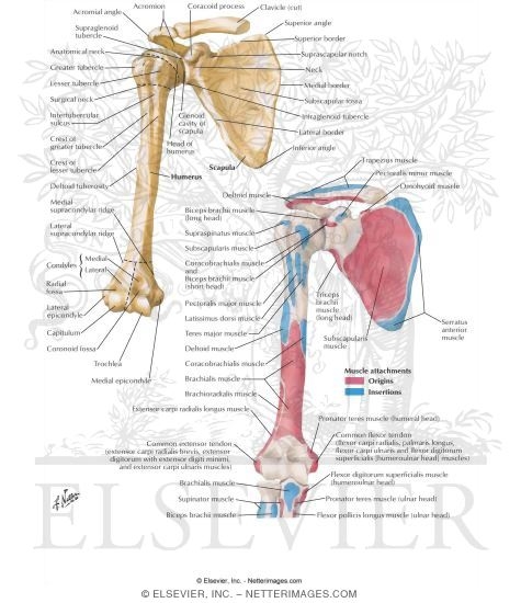
SCAPULA
1-Scapula:-In anatomy,
the
scapula (plural
scapulae) (Medical Latin), or
shoulder
blade, is the
bone
that connects the
humerus
(upper arm bone) with the
clavicle (collar bone).
The scapula forms the posterior (back) located part of the
shoulder
girdle. In
humans,
it is a flat bone, roughly
triangular in shape, placed on a posterolateral aspect of
the
thoracic
cage.
SOME
IMP POINTS:-
1-NOTCH:
notch (
noch)
incisure; an indentation(A notch, a pit, or a depression) on the edge of a bone
or other organ.
2-
Fossa (
/ˈfɒsə/;
[1][2]
plural
fossas /ˈfɒsəz/, or
fossae (
/ˈfɒsiː/ or
/ˈfɒsaɪ/); from the Latin "
fossa",
ditch or trench) is a word used in anatomical nomenclature to describe a depression
or hollow usually in a bone.
PARTS OF SCAPULA:-
1-The
glenoid cavity (or
glenoid fossa of scapula from
Greek:
gléne, "socket") is
a part of the
shoulder.
It is a shallow
pyriform,
articular
surface, which is located on the lateral angle of the
scapula. It is
directed laterally and forward and articulates with the head of the
humerus; it is
broader below than above and its vertical diameter is the longest.
This cavity forms the
glenohumeral joint along with the
humerus. This
type of joint is classified as a
synovial,
ball and socket joint.
2-The
coracoid process (from
Greek
κόραξ, crow) is a small hook-like structure on the lateral edge of the superior
anterior portion of the
scapula. Pointing laterally forward, it, together with the
acromion,
serves to stabilize the
shoulder joint. It is
palpable in
the
deltopectoral groove between the
deltoid
and
pectoralis major muscles.
"Coracoid" in itself means "like a
raven's beak",
with reference to its shape. (Greek "Korax" = Raven)
3-In human anatomy, the
acromion (from Greek:
akros,
"highest",
ōmos, "shoulder", plural: acromia) is a
bony process on the
scapula (shoulder blade), together with the
coracoid
process extending laterally over the
shoulder joint. The acromion is a continuation
of the
scapular spine, and hooks over anteriorly. It
articulates with the
clavicle (collar bone) to form the
acromioclavicular joint.
4-The suprascapular notch (or scapular notch) is a notch in
the superior border of the scapula, just medial to the base of the
coracoid
process.
This notch is converted into a
foramen by the
superior transverse scapular
ligament(The
superior transverse ligament (
transverse or
suprascapular
ligament) converts the
scapular notch into a foramen or opening.) and
serves for the passage of the
suprascapular nerve.
5-
subscapular fossa.
The costal or ventral surface of the
scapula presents
a broad concavity, the
subscapular fossa.

It provides an attachment for the
subscapularis muscle(The
subscapularis
is a large triangular muscle which fills the
subscapular
fossa and inserts into the
lesser
tubercle of the
humerus and the front of the capsule of the shoulder-joint.)
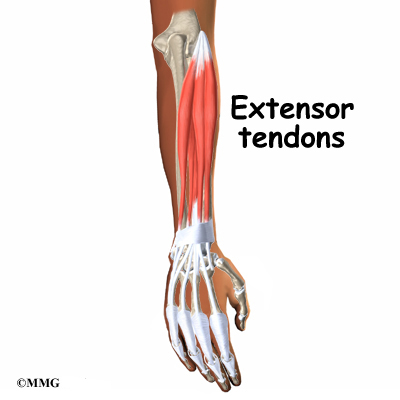
MUSCLES:-
Note:- Muscles mentioned in scapula
are not repeated:-
1-Omohyoid Muscle:- The omohyoid muscle is a muscle that
depresses the
hyoid.
It is located at the front of the
neck and consists of two bellies separated by an intermediate
tendon.
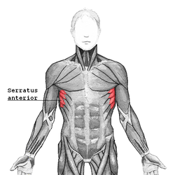
2-Serratus anterior muscle:- The
serratus anterior ( Latin: serrare =
to saw, referring to the shape, anterior = on the front side (of the body)) is
a muscle that originates on the surface of the second to ninth ribs at the side
of the chest and inserts along the entire anterior length of the medial border
of the
scapula.
3-
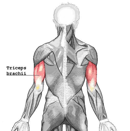
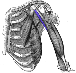
triceps brachii muscle :-The
triceps brachii muscle (
Latin for
"three-headed arm muscle") is the large
muscle on the
back of the
upper limb
of many
vertebrates.
It is the muscle principally responsible for
extension of the
elbow joint
(straightening of the arm).
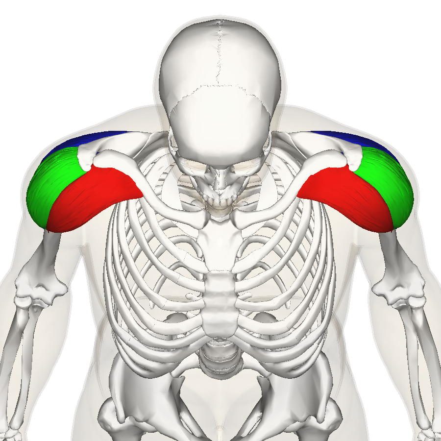
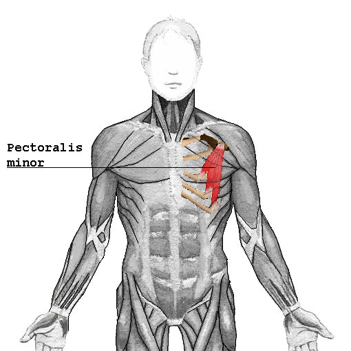
4- Pectoralis minor:-The
pectoralis minor is a thin, triangular
muscle, situated at the upper part of the
chest, beneath the
pectoralis
major in the human body.
6- In
human anatomy, the
biceps brachii, or simply
biceps
in common parlance, is, as the name implies, a two-headed
muscle. The biceps
lies on the upper arm between the shoulder and the elbow. Both heads arise on
the
scapula
and join to form a single muscle belly which is attached to the upper forearm.
While the biceps crosses both the
shoulder and
elbow joints, its main function is at the latter where it flexes the elbow and
supinates
the forearm
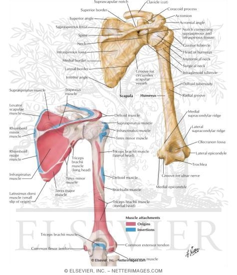
POSTERIOR
VIEW:-
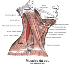
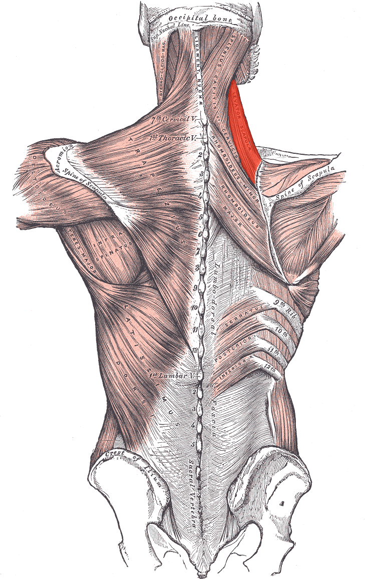
1-In human anatomy, the
levator scapulae is a
skeletal
muscle situated at the back and side of the neck. As the name suggests, its
main function is to lift the
scapula.
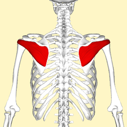
2-In human anatomy, the
infraspinatus muscle is a thick triangular
muscle, which
occupies the chief part of the
infraspinatous fossa.
[1]
As one of the four muscles of the
rotator
cuff, the main function of the infraspinatus is to externally rotate the
arm and stabilize the shoulder joint.
3-The
latissimus dorsi (plural:
latissimi dorsi), meaning
'broadest [muscle] of the back' (
Latin latus meaning 'broad',
latissimus meaning
'broadest' and
dorsum meaning the back), is the larger, flat,
dorso-lateral muscle on the trunk, posterior to the arm, and partly covered by
the
trapezius
on its median dorsal region.
Latissimi dorsi are commonly known as
"
lats", especially among
bodybuilders.
The latissimus dorsi is responsible for
extension,
adduction,
transverse extension also known as horizontal abduction, flexion from an
extended position, and (medial)
internal
rotation of the
shoulder joint. It also has a
synergistic
role in extension and lateral flexion of the lumbar spine.
Due to bypassing the scapulothoracic joints and attaching
directly to the spine, the actions the latissimi dorsi have on moving the arms
can also influence the movement of the scapulae, such as their downward
rotation during a
pull up
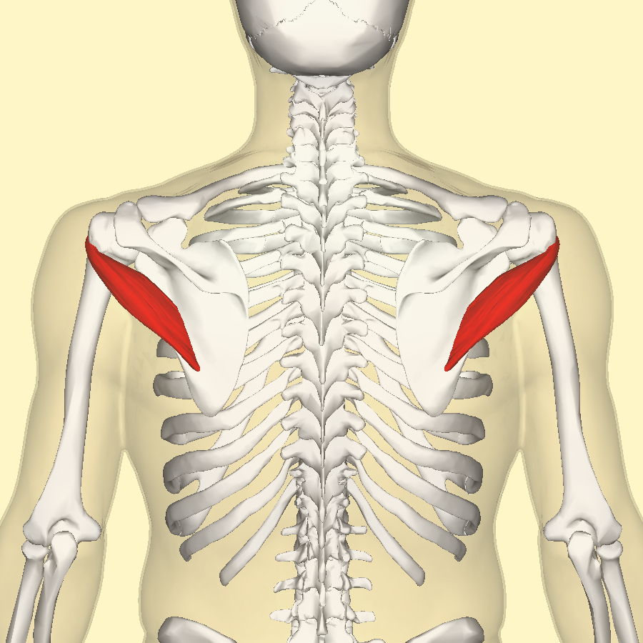
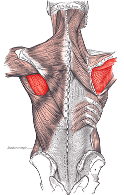
4-The
teres major muscle is a muscle of the
upper limb
and one of six scapulohumeral muscles. It is a thick but somewhat flattened
muscle, innervated by the
lower subscapular nerve (c5,c6).
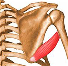
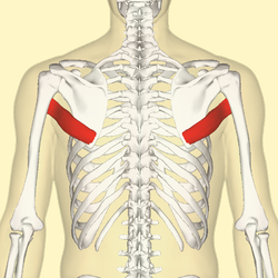
5-The
teres minor (
Latin teres
meaning 'rounded') is a narrow, elongated muscle of the
rotator
cuff
6-The
rhomboid major is a
skeletal
muscle on the back that connects the
scapula with the
vertebrae of
the
spinal
column. In human anatomy, it acts together with the
rhomboid minor to keep the scapula pressed
against thoracic wall and to retract the scapula toward the vertebral column.
[1]
7-In human anatomy, the
rhomboid minor is a small
skeletal
muscle on the back that connects the
scapula with the
vertebrae of the
spinal column.
Located inferior to
levator scapulae and superior to
rhomboid major, it acts together with the
latter to keep the scapula pressed against the thoracic wall. It lies deep to
trapezius
but superficial to the long spinal muscles
 (NOTE:-ABOVE INFORMATION MIGHT HAVE ERRORS,USE AT YOUR OWN RESPONSIBILITY)


 (NOTE:-ABOVE INFORMATION MIGHT HAVE ERRORS,USE AT YOUR OWN RESPONSIBILITY)